led by
Dr. Thibault Cholvin
Using in vivo two-photon calcium imaging in behaving mice, we explore how the brain captures and ensures long-term storage of experiences in cortico-hippocampal networks.
Research
We conduct research on spatial navigation and episodic memory in behaving mice. Using two-photon in vivo calcium imaging to track the activity of neurons over extended periods of time (multiple days / weeks), we shed light on the properties of the different subregions of the hippocampus (CA1, CA3, DG) as well as their cortical inputs (such as the axonal projections coming from the entorhinal cortex). For this, we combine two-photon imaging and virtual reality to simultaneously capture the activity of hundreds of neurons or axons while the mouse is navigating through virtual environments. Finally, we use the recorded cell or axon activity to decode the animals’ concurrent location and environment, and thus infer the relative involvement of these brain regions in spatial memory.
Techniques
in vivo two-photon Calcium imaging
We perform two-photon calcium imaging in head-fixed mice performing goal-oriented tasks in familiar or virtual environments.
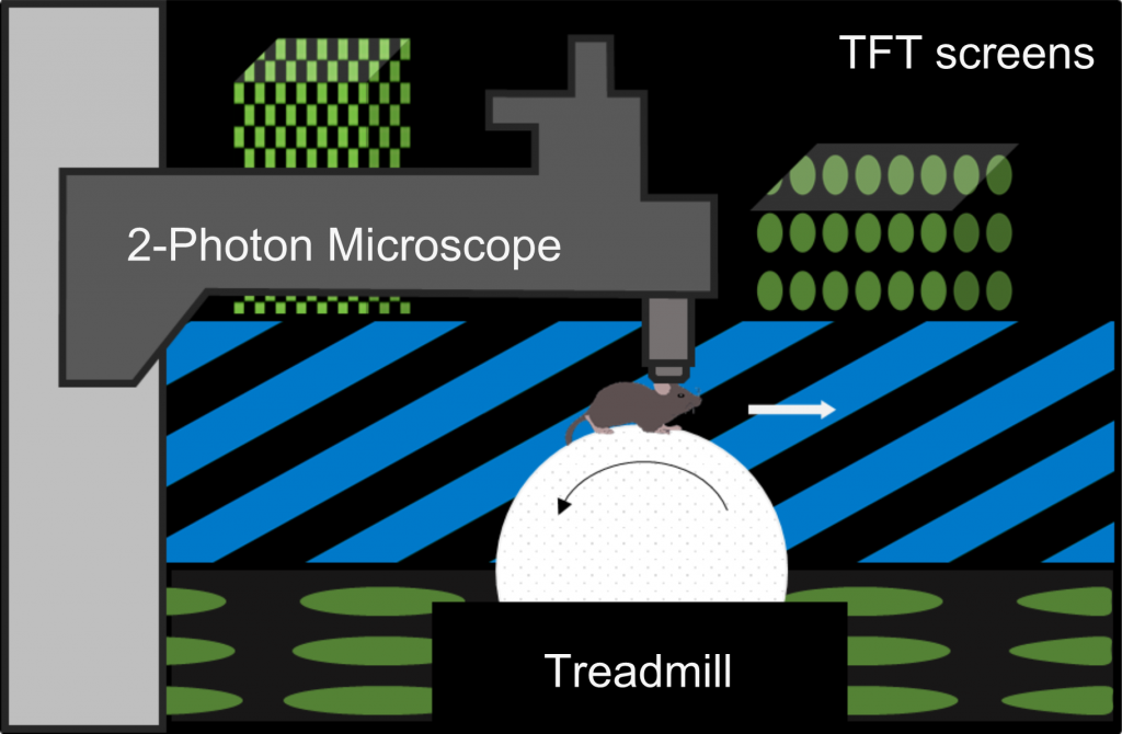
The animal is standing on an air-supported polystyrene ball, allowing it to navigate through virtual worlds while keeping its actual position constant.
Screens arranged in a hexagonal arc around the mouse and placed ~25 cm away from the head of the animal cover ~260° of the horizontal and ~60° of the vertical visual field of the mouse.

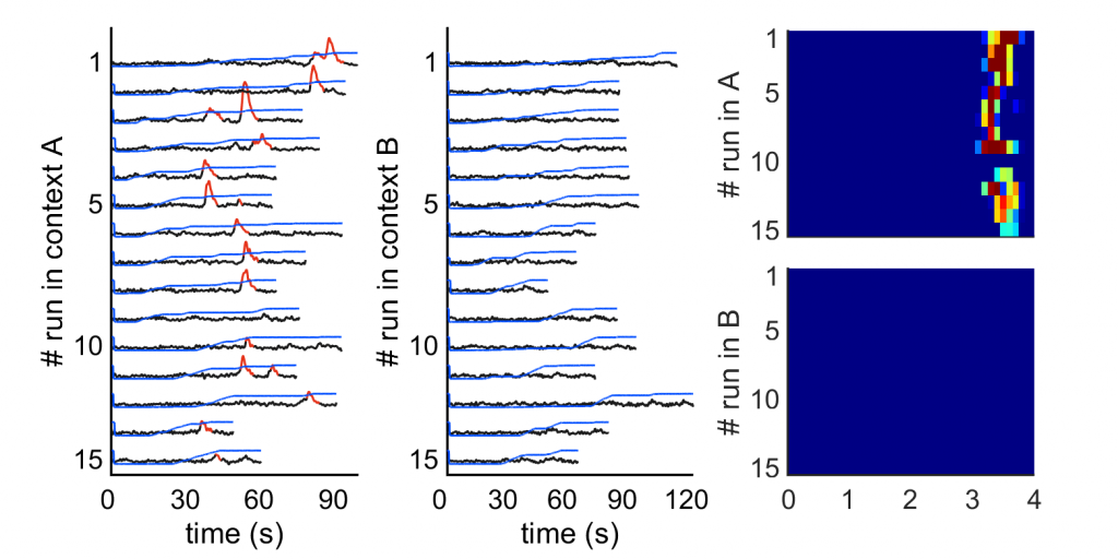
We use in vivo two-photon calcium imaging to simultaneously capture the activity of hundreds of cells in one or the other subregions of the hippocampus. In this example, we panneuronally expressed GCaMP in the hippocampus, allowing us to image the pyramidal cells in CA1 and CA3 as well as the granule cells of the dentate gyrus.

The traces below are showing the activity of a single granule cell over 15 runs in two different environments; note the reliable presence of a place field in context A, as opposed to the striking absence of activity of this cell in context B.
This video shows a typical example of a field of view encompassing a large population of granule cells that we can image in our experiments.
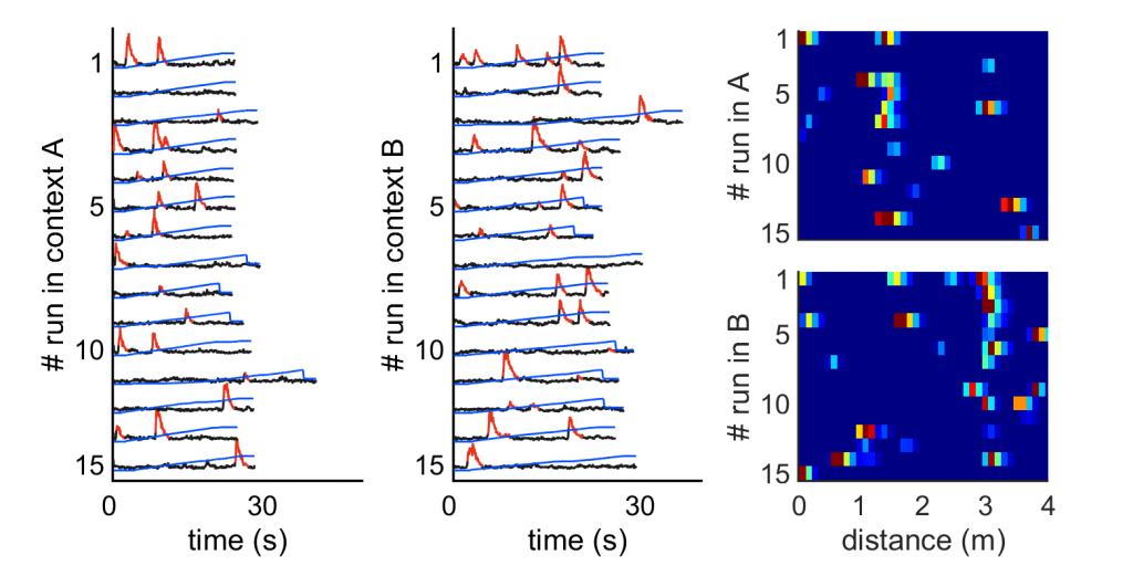
In vivo two-photon calcium imaging can also be applied to axonal projections, allowing us to capture the activity of hundreds of axon terminals coming from neurons located in distant brain areas (such as the entorhinal cortex) and projecting to the hippocampus.

The traces below are showing the activity of a single bouton (axon terminal) over 15 runs in two different environments; note the presence of place fields in both contexts, but in distinct locations depending on the context (remapping).
This video shows a typical example of the field of view encompassing numerous axonal projections from the MEC to the dentate gyrus (middle molecular layer) as we can image them in our experiments.
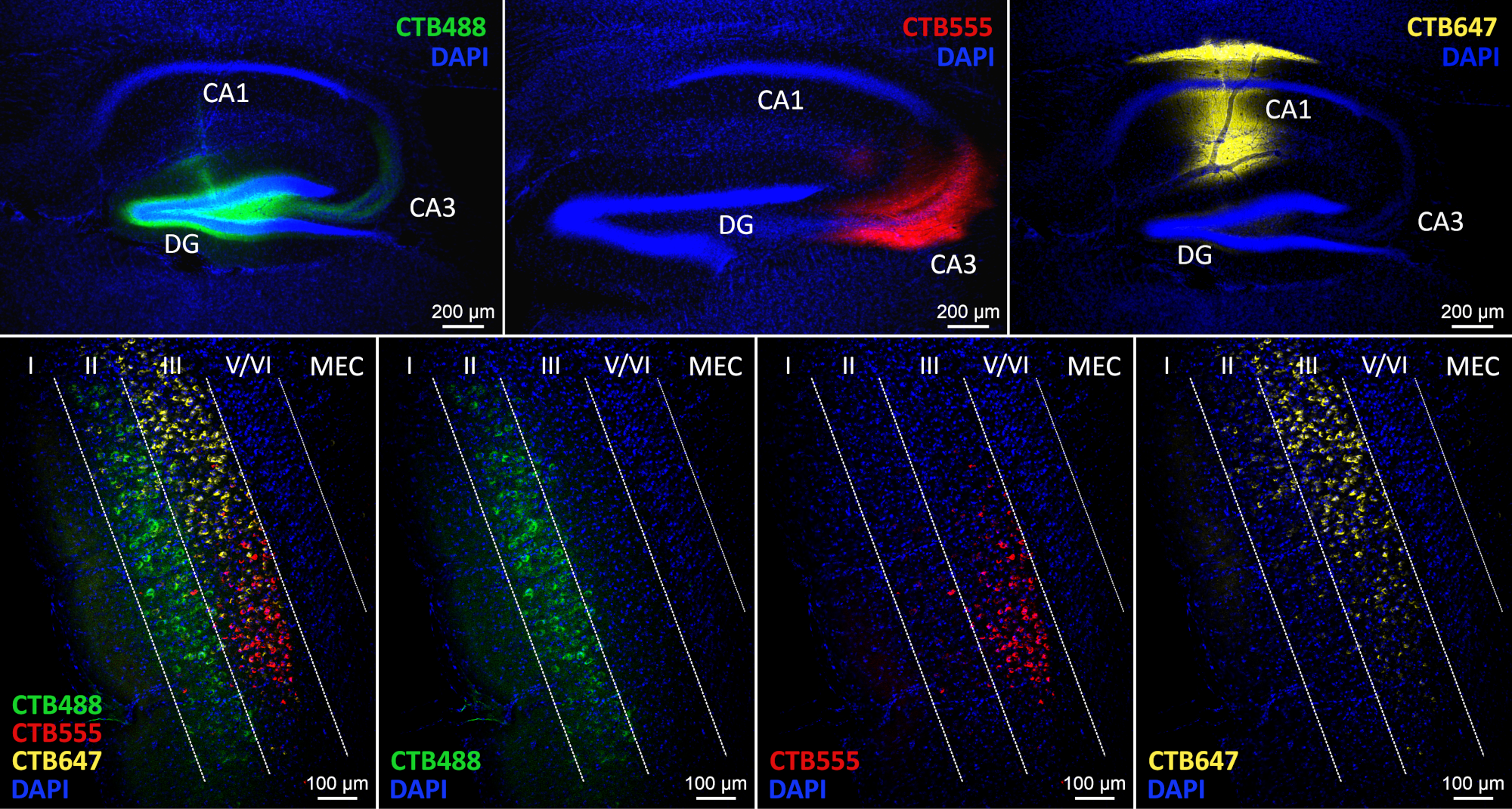
Using retrograde tracers such as cholera toxin subunit B (CTB) conjugated with different fluorophores (e.g. Alexa Fluor 488 (green), 555 (red) or 647 (blue)), we can examine multiple neuronal pathways within the same brain, for example the projections from the entorhinal cortex to specific subregions of the hippocampus. In the example presented here, 3 different variants of CTB have been injected in CA1, CA3 and the DG, respectively, in a single animal, revealing the differences (e.g. layer specificity) in the connectivity between the entorhinal cortex and each of the subregions of the hippocampus.
Chemogenetic tools such as designer receptors exclusively activated by designer drugs (a.k.a. DREADDs,) allow for the remote control of neuronal activity. DREADDs are engineered muscarinic or opioid receptors that remain silent until triggered by specific ligands (that can easily be administrated via a simple I.P. injection). Depending on their type, DREADD receptors can either induce an increase (hM3Dq) or decrease (hM4Di) of neuronal activity.
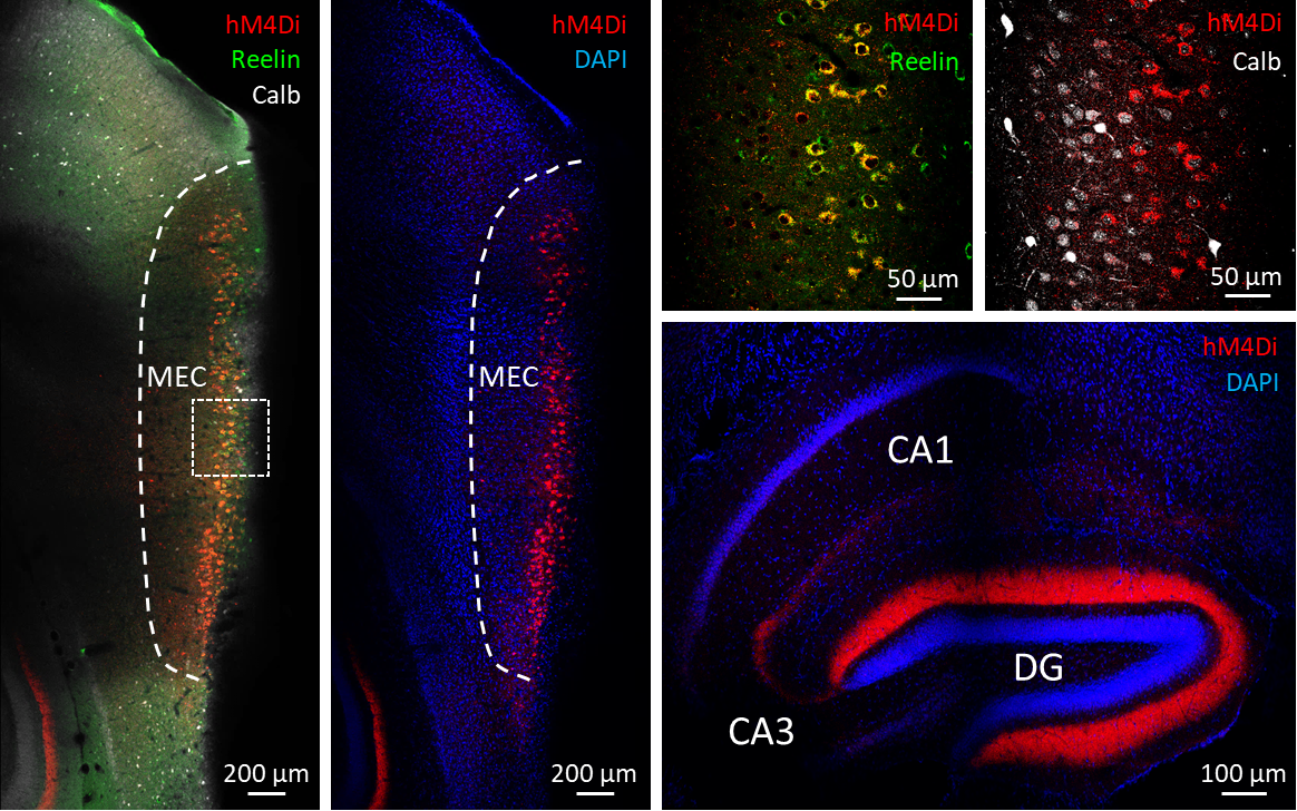
In this example, activity of the stellate cells of the MEC can be down-regulated through the expression of hM4Di receptors, giving us the opportunity to assess the importance of the inputs coming from this cell population to the hippocampus on hippocampal neurons activity.
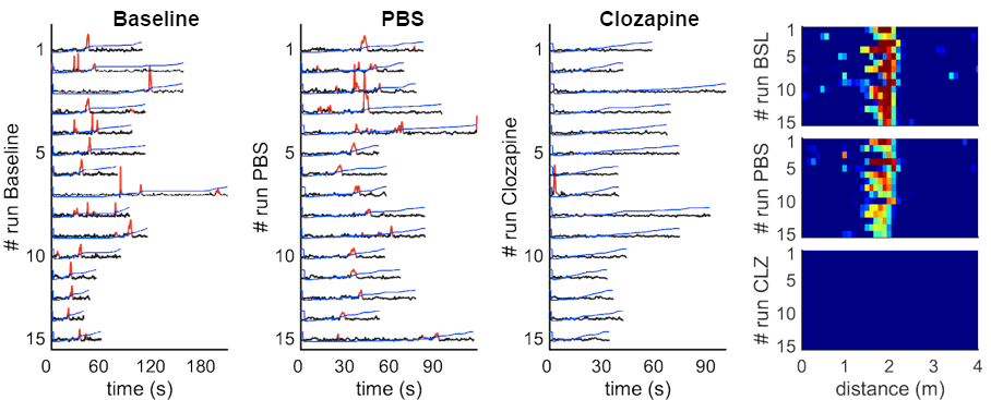
The traces below are showing the activity of a single granule cell of the dentate gyrus recorded under baseline, PBS and Clozapine conditions. Following activation of the hMD4i receptors expressed by the MEC layer-2 stellate cells using Clozapine, this granule cell is silenced.
To directly address how different neuronal populations are participating in the encoding of space and context, we use an inverse approach, starting from the recorded cell (or bouton) activity to decode the animals’ concurrent location and context (using a population-vector-based method).
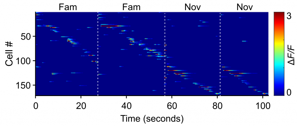
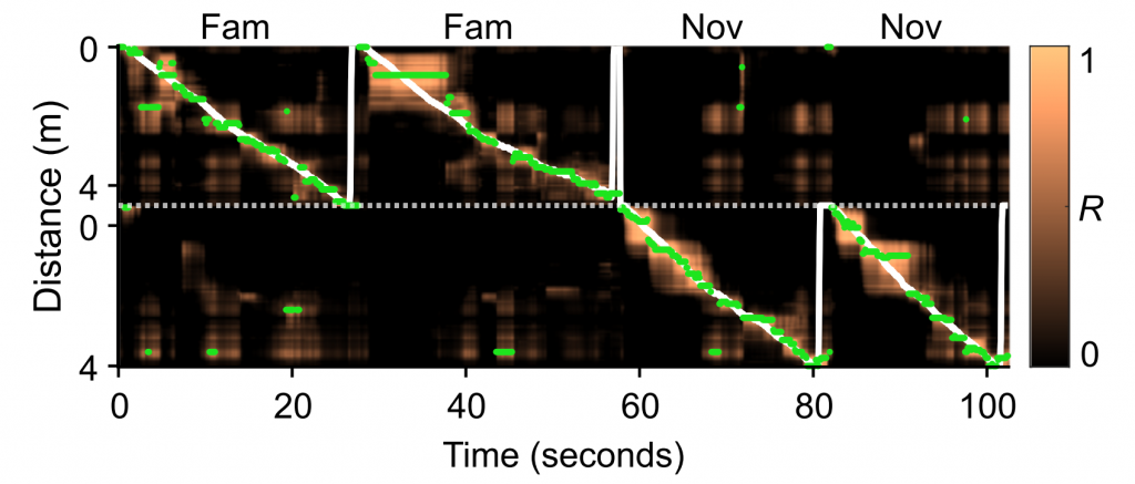
In the example presented here, we randomly selected subsamples of 50 cells from all recorded cells in a given session, either from left or right hippocampi.
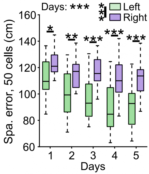
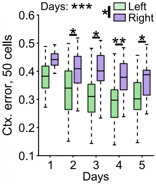
Decoding performance for both space and context increased over days in both left and right granule cells populations. Nevertheless, decoding errors were always substantially smaller for left granule cell populations, suggesting that contextual information is differentially processed by granule cells depending on the brain hemisphere.
Team members
Publications
Hemisphere-specific spatial representation by hippocampal granule cells. Cholvin T, Bartos M. Nature Commun. 2022 Oct 20;13(1):6227.
The hippocampus converts dynamic entorhinal inputs into stable spatial maps. Cholvin T, Hainmueller T, Bartos M. Neuron. 2021, 109:3135-3148.e7.
Ventral midline thalamus lesion prevents persistence of new (learning-triggered) hippocampal spines, delayed neocortical spinogenesis, and spatial memory durability. Klein MM, Cholvin T, Cosquer B, Salvadori A, Le Mero J, Kourouma L, Boutillier AL, Pereira de Vasconcelos A, Cassel JC. Brain Struct Funct. 2019, 224:1659-1676.
Ventral midline thalamus is necessary for hippocampal place field stability and cell firing modulation. Cholvin T, Hok V, Chaillan FA, Giorgi L, and Poucet B. J Neurosci. 2018, 38:158-172.
Using MRI to predict the fate of excitotoxic lesions in Rats. Cholvin T, Giorgi L, Baril N, Brezun JM, Poucet B, and Chaillan FA. PLoS One. 2018, 13:e0200659.
Environmental enrichment enhances systems-level consolidation of a spatial memory after lesions of the ventral midline thalamus. Ali M, Cholvin T, Muller MA, Cosquer B, Kelche C, Cassel JC, Pereira de Vasconcelos A. Neurobiol Learn Mem. 2017, 141:108-123.
Dorsal hippocampus and medial prefrontal cortex each contribute to the retrieval of a recent spatial memory in rats. Cholvin T, Loureiro M, Cassel R, Cosquer B, Herbeaux K, de Vasconcelos AP, Cassel JC. Brain Struct Funct. 2016, 221:91-102.
The ventral midline thalamus contributes to strategy shifting in a memory task requiring both prefrontal cortical and hippocampal functions. Cholvin T, Loureiro M, Cassel R, Cosquer B, Geiger K, De Sa Nogueira D, Raingard H, Robelin L, Kelche C, Pereira de Vasconcelos A, Cassel JC. J Neurosci. 2013, 33:8772-8783.
The ventral midline thalamus (reuniens and rhomboid nuclei) contributes to the persistence of spatial memory in rats. Loureiro M, Cholvin T, Lopez J, Merienne N, Latreche A, Cosquer B, Geiger K, Kelche C, Cassel JC, Pereira de Vasconcelos A. J Neurosci. 2012, 32:9947-9959.
- The reuniens and rhomboid nuclei of the thalamus: A crossroads for cognition-relevant information processing? Cassel JC, Ferraris M, Quilichini P, Cholvin T, Boch L, Stephan A, Pereira de Vasconcelos A. Neurosci Biobehav Rev. 2021, 126:338-360.
- The reuniens and rhomboid nuclei: neuroanatomy, electrophysiological characteristics and behavioral implications. Cassel JC, Pereira de Vasconcelos A, Loureiro M, Cholvin T, Dalrymple-Alford JC, Vertes RP. Prog Neurobiol. 2013, 111:34-52 (Review).
